Treatment
Uterine Fibroids
Uterine fibroids, also known as uterine leiomyomas, fibromyoma or fibroids, are noncancerous smooth muscle tumor. These tumors rarely becomes cancer (0.1%)..
Fibroids grow in the muscle layer of your uterus (womb). Fibroids are very common, they vary in size, from being tiny to a very large size like that of a melon.
Some patient present with a single fibroid whereas others can have multiple fibroids varying in sizes
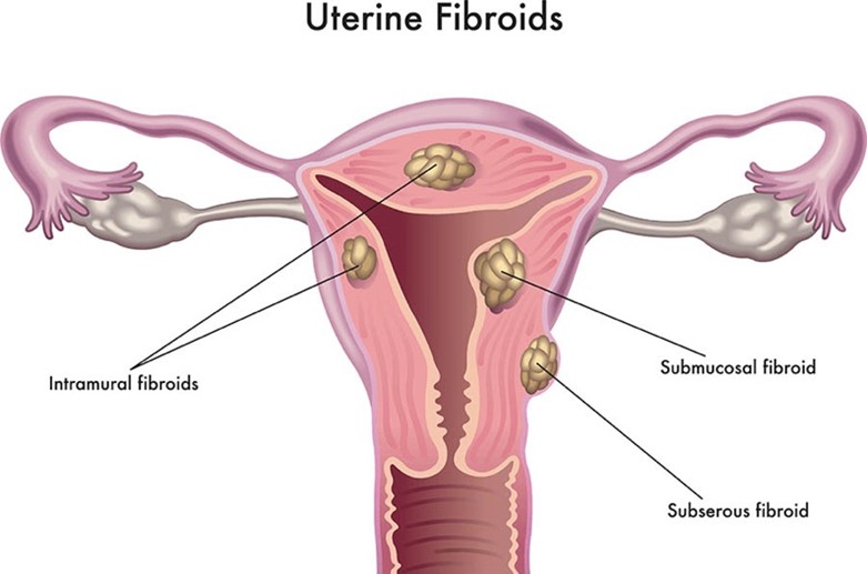
There are different types of fibroids, based on where they grow in uterus.
- Subserous or subserosal fibroids grow on the outer wall of the uterus
- Intramural fibroids grow within the muscle wall of uterus.
- Submucosal fibroids grow on the inner wall of uterus.
- Pedunculated fibroids are a type of fibroid that are attached to uterus with a stalk.
Symptoms
If the fibroids are small, they do not cause any symptoms are encountered incidentally during health check ups. If fibroids are symptomatic then patient may present with
- heavy periods
- Prolonged bleeding
- pain during periods
Other symptoms can include:
- Lower abdominal pain or pressure symptoms
- painful sex
- needing to urinate frequently
Symptoms from fibroids usually get better after menopause.
Causes
The cause of uterine fibroids is unknown. However, the female hormones like oestrogen and progesterone are responsible for the growth of fibroids.
Fibroids usually develop during reproductive years. They may shrink after menopause.
Diagnosis
To diagnose uterine fibroids, one of the following tests can be performed
History and Physical examination
Tests that can help diagnose fibroids include:
- Pelvic Ultrasound(Transabdominal and Transvaginal ultrasound) – A procedure during which a small instrument, called a transducer, is either inserted into the vagina or pressed over the abdomen to produce pictures of the internal organs using sound waves. The doctor can see the size, shape and texture of the uterus and evaluate any growths.
- Magnetic resonance imaging (MRI) – This is a form of advanced imaging technology that provides highly detailed images of internal organs. These images help your provider determine the exact location and characteristics of fibroids and, if needed, plan minimally invasive treatments.
- Hysterosalpingography – This is a type of X-ray exam of the uterus and fallopian tubes. Your doctor will use a special dye to more easily visualize these organs and determine if the fibroids have blocked your fallopian tubes.
- Hysteroscopy – This is a visual exam of the canal of the cervix and the interior of the uterus using a viewing instrument (hysteroscope) inserted through the vagina.
- Laparoscopy (keyhole surgery) – where a thin telescope is inserted through a small cut in abdomen (tummy) to look at reproductive organs
Treatment
Uterine fibroids don’t always need to be treated. Treatment may be recommended if
- patient is having symptoms
If treatment is recommended, the type of treatment will depend on:
- symptoms
- the type, size and number of fibroids
- age of the patient
- whether patient desire future pregnancy or wish to preserve uterus
There are several different treatment options.
Medical treatments
Medicines and hormone treatments can be given to:
- shrink the size of fibroid
- treat symptoms
They can include medicines, implants and intra-uterine devices.
Procedures or surgery
Surgery or other procedures can be done to shrink or remove your fibroids. These include:
- MRI-guided focused ultrasound (HIFU) – A non-invasive technique that uses sound waves to destroy fibroids.
- Uterine artery embolization (UAE) – A minimally invasive procedure that blocks blood supply to fibroids, causing them to shrink.
- Surgical removal of fibroids, called a myomectomy. This can be done via two route – hysteroscopy or laparoscopy.
- Hysterectomy – Surgical removal of the uterus, a permanent solution for fibroids.